Monitoring Cell Culture Inside an Incubator with the ioLight Digital Microscope
Monitoring cell cultures regularly is an important part of many laboratory workflows, helping researchers assess cell growth, morphology, confluence and overall culture health. However, traditional cell culture monitoring often requires samples to be removed from their controlled environment and transferred to a microscope.
For sensitive or long-running experiments, repeatedly moving cultures between an incubator and microscope can be less than ideal. Opening the incubator can also temporarily alter the carefully controlled temperature, humidity and CO₂ conditions that cells rely on.
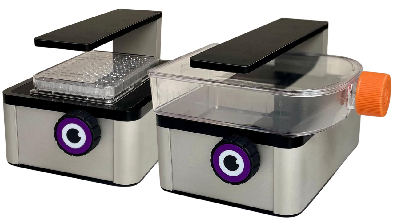
An incubator microscope offers a different approach. By placing a compact microscope directly inside the incubator, researchers can monitor cultures while they remain in their controlled environment. The ioLight Compact Cell Imager has been designed specifically to provide this type of live cell imaging, allowing researchers to view and record cell cultures without repeatedly opening the incubator.
Why monitor cell cultures inside the incubator?
Cell cultures can change considerably over the course of an experiment. Regular observation can help researchers understand how cells are developing and identify changes that may require attention.
With a conventional microscope, cultures generally need to be removed from the incubator before they can be examined. This can introduce unnecessary handling and temporarily expose the cultures to conditions outside their controlled environment.
An incubator microscope for cell culture allows images to be captured while the culture remains inside the incubator. This can be particularly useful for long-running experiments, delicate cultures and applications where maintaining stable environmental conditions is important.
The ioLight Compact Cell Imager can be left inside a CO₂ incubator and transmits images wirelessly to a smartphone, tablet or computer outside the chamber. This means researchers can view their cultures without opening the incubator door.
The benefits of live cell imaging inside an incubator
1. Monitor cells without opening the incubator
One of the key benefits of an incubator microscope is the ability to observe cell cultures without removing them from their controlled environment.
The ioLight microscope sits inside the incubator, while the display remains outside. Images can be viewed on a compatible smartphone, tablet or computer, allowing researchers to check their cultures without opening the incubator door.
This can help reduce unnecessary disturbance to the culture and makes it easier to carry out more frequent observations.
2. Observe cell growth and morphology
Live cell imaging can provide useful information about the condition and development of a culture.
The ioLight Compact Cell Imager can be used to observe features including cell number, morphology, motility and confluence. This can give researchers and laboratory technicians a clearer picture of how a culture is progressing between routine checks.
For example, monitoring changes in cell morphology or confluence over time can help researchers assess whether a culture is developing as expected.
3. Support earlier identification of potential problems
Regular cell culture monitoring can also help researchers identify changes at an earlier stage.
Unexpected changes in cell appearance, growth or behaviour may indicate that a culture requires further investigation. The ability to observe cultures more frequently, without repeatedly removing them from the incubator, can therefore provide additional information when assessing cell health and experimental progress.
ioLight highlights applications including monitoring HEK-293, CHO and HeLa cells, as well as identifying contaminants and assessing cell health.
4. Capture time-lapse images
Some cell culture experiments require researchers to understand how cells change over hours or days rather than relying on individual observations.
The ioLight Compact Cell Imager supports time-lapse imaging, allowing images to be captured at regular intervals. This can create a visual record of changes such as cell growth and development over the course of an experiment.
Time-lapse imaging can be particularly useful when studying dynamic processes where observing the progression of an experiment is just as important as the final result.
5. Reduce unnecessary sample handling
Moving cultures between an incubator and microscope introduces additional handling steps into a workflow.
By providing live cell imaging inside the incubator, an incubator microscope can reduce the need to repeatedly move samples for routine visual checks. This can be beneficial when working with delicate cultures or experiments that need to remain undisturbed for extended periods.
The ioLight can also be used for applications such as wound healing assays, chemotaxis and delicate 3D cultures including organoids, where maintaining a stable environment can be particularly important.
A compact microscope designed for incubator use
Space can be another consideration when introducing additional equipment into a laboratory incubator.
The ioLight Compact Cell Imager has a small footprint and is designed to operate inside humid CO₂ environments. Its sealed construction allows it to remain inside an incubator and be sterilised after use.
Samples can be viewed from below in Petri dishes, flasks, well plates and cavity slides, providing flexibility for different cell culture workflows. The standard Compact Cell Imager provides approximately 1 µm resolution with a field of view of around 1.2 × 0.9 mm.
The ioLight Compact Cell Imager range
The ioLight range includes three Compact Cell Imager models, allowing users to select the imaging capabilities most appropriate for their application.
Compact Cell Imager
The standard Compact Cell Imager provides a range of imaging modes for live cell imaging and routine cell culture monitoring.
Key features include:
- Brightfield imaging
- Low NA and high-contrast brightfield
- Darkfield imaging
- Digital phase contrast
- Approximately 1 µm resolution
- Time-lapse imaging
- Z-stack imaging
- Wireless image display on a smartphone, tablet or computer
- Suitable for Petri dishes, flasks and well plates
- Designed for use inside CO₂ incubators, safety cabinets and other controlled environments
Z-stack imaging can be particularly useful for larger or three-dimensional samples, including organoids and embryos.
Compact Cell Imager with Fluorescence
For applications requiring fluorescence imaging, the Compact Cell Imager with Fluorescence combines the imaging capabilities of the standard model with epifluorescence.
It can detect a range of commonly used fluorophores, including GFP, YFP, propidium iodide (PI), SYTO9 and DS Red. The system retains brightfield, darkfield, high-contrast and digital phase contrast imaging, providing researchers with multiple imaging options within a single compact system.
The fluorescence model is therefore suited to applications where researchers need to combine conventional live cell imaging with fluorescence-based observations.
Compact Cell Imager with Dual Band Fluorescence
The Compact Cell Imager with Dual Band Fluorescence provides brightfield and dual-band fluorescence imaging for applications requiring a broader range of fluorescent markers.
Supported fluorophores include GFP, YFP, PI, SYTO9, DS Red, tdTomato, Alexa Fluor, FITC and mCherry, amongst others.
This makes the dual-band model an option for more advanced fluorescence applications where monitoring multiple fluorescent markers is important.
Bringing microscopy closer to the experiment
An incubator microscope changes the way researchers can approach cell culture monitoring. Instead of periodically removing cultures for observation, the microscope can remain with the experiment, providing access to images while the cells remain inside their controlled environment.
For routine cell culture monitoring, this can provide a convenient way to observe cell growth, morphology, confluence and motility. For longer experiments, time-lapse imaging can provide a record of how cultures change over time, while fluorescence models add another level of information for applications using fluorescent markers.
The compact design of the ioLight Compact Cell Imager also means it can be introduced into existing laboratory workflows without requiring a large conventional microscope inside the incubator.
Find the right ioLight cell culture microscope for your application
From routine brightfield and digital phase contrast imaging through to fluorescence and time-lapse applications, the ioLight Compact Cell Imager range provides flexible options for live cell imaging and cell culture monitoring inside an incubator.
If you are looking for a digital microscope for cell culture, an incubator microscope for use with a CO₂ incubator, or a compact system for time-lapse live cell imaging, the ioLight range offers a practical solution for monitoring cultures while they remain in their controlled environment.
You can see the full range of iolight products HERE.
